Understanding Preclinical Research at Neuralink
Introduction
Preclinical research, also called nonclinical testing, refers to the body of work and scientific studies that occur before any new product (device or drug) is tested in humans (at “the clinic”). The FDA Modernization Act 2.0 defines nonclinical testing as, “a test conducted in vitro, in silico, or in chemico, or a non-human in vivo test that occurs before or during the clinical trial phase of the investigation of the safety and effectiveness of a drug, and may include animal tests, or non-animal or human biology-based test methods, such as cell-based assays, microphysiological systems, or bioprinted or computer models.”
Generally, preclinical research can be split into two major categories: in vitro and in vivo. All tests conducted in a controlled environment outside of a living organism (think cell culture, test tubes, and lab bench work) are considered in vitro research — Latin for “within the glass.” Any tests that are conducted within a living organism (plant, animal, or human) are considered in vivo research — Latin for “within the living.”
At Neuralink, preclinical studies are further subdivided into four categories: bench, pilot, research and development (R&D), and good laboratory practices (GLP).
Bench
Bench studies are the first step taken towards evaluating a scientific or engineering idea. They serve as a “proof of concept” validation or rejection phase — focusing on the basic mechanical, chemical, proxy, or in vitro tests in order to narrow down the research focus and identify variables.
Pilot
Pilot studies are the second step in the preclinical research pipeline. These tests are iterative and are performed in order to demonstrate that the new idea/method/design is compatible with the current standard of practice, results in promising initial data, and is logistically feasible.
R&D
R&D studies are the third step in this process and are critical to establishing readiness for GLP. These studies are designed to fine-tune the study hypotheses, objectives, methods, and metrics.
GLP
GLP studies are the final step in the preclinical research process and focus primarily on the evaluation of the safety and effectiveness of a device and/or any new parts or materials that will be in contact with patient tissue. GLP studies are conducted per the Code of Federal Regulations Title 21, Part 58 as defined by the FDA and are the highest industry standard.
These studies are precursors to clinical trials and the FDA approves preliminary clinical trials based on the results from GLP studies. Because of this, companies are subject to inspections and audits from the FDA to ensure compliance with these federal regulations.
Preclinical Research at Neuralink
The preclinical team is responsible for the design, conduct, documentation, analysis, and reporting of preclinical studies. It is our responsibility to provide a means for ensuring the implantation of Neuralink devices into animals, and ultimately humans, is safe, effective, and robust.
Currently, the team is composed of neuroscientists, neuro-engineers, biomedical engineers, and veterinarians. Preclinical team conduct is guided by prioritizing two main tenets:
- Animal Welfare
- Scientific Rigor
Before any new study, method, procedure, or approach is initialized, the preclinical team works alongside the Institutional Animal Care and Use Committee (IACUC) to align on best practices with regards to the welfare of the animal. Neuralink’s IACUC has a multi-step process to review, deliberate, and vote on study protocols and methods. Approval by the IACUC is required prior to initiating any animal study at Neuralink.
The cross-functional collaboration at Neuralink between the preclinical, veterinary medicine, animal care, pathology, engineering, IACUC, and surgical teams provides a balanced and fair perspective on study design that then enables the team to develop the most comprehensive protocols possible.
There is no single animal model that perfectly recapitulates human anatomy and physiology. We employ a variety of models for different research purposes. Below you will find a table that outlines the benefits and drawbacks of using each of our animal models. The model chosen for any specific study is based on the objectives, a risk-benefit analysis, and the likelihood of representing the human condition under study. Whenever possible, at the end of their research involvement, Neuralink will seek ethical retirement options for all of our species that allow them to live out their lives in sanctuaries and on farms.
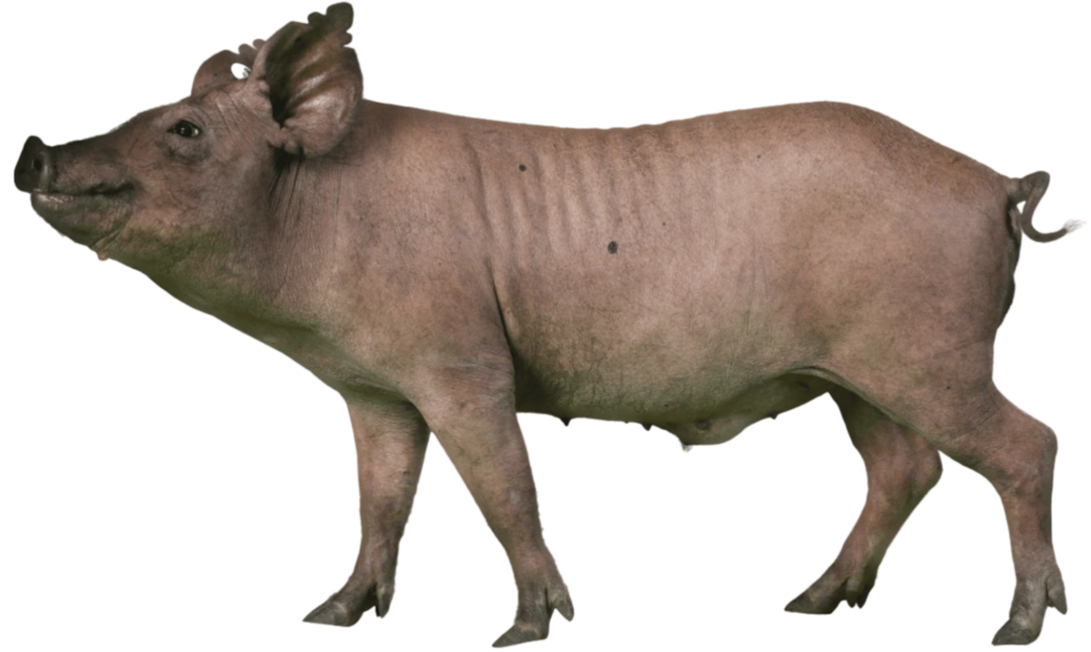
Porcine
(Pig)
Pigs are well suited for studies that closely mimic the human condition in terms of brain size, skull thickness, and recovery time. Pigs are also able to quickly learn a variety of behaviors utilizing positive reinforcement, such as simple discrimination tasks and voluntary hoof trims. One reason to consider an animal model other than swine is due to the biologically normal expansion of the frontal sinuses over time, along with the rate and direction of skull growth.
Ovine
(Sheep)
Sheep are an excellent, non-primate representation of the human condition. Along with the rate of their skull and neomembrane regrowth, sheep also have more natural brain motion than pigs, which makes them the better model for representing surgical conditions in people. Sheep are also conditioned to voluntarily participate in necessary husbandry and medical behaviors such as shearing and stationing for veterinary assessments. One reason to consider an animal model other than sheep is due to challenges with ruminant metabolism during prolonged anesthesia.
Non-Human Primate
(Monkey)
Monkeys are the closest available relative to humans and, for that reason, are the best functional approximation to the human condition in a research setting. Monkeys are well suited for studies that involve the complex behavioral paradigms required to assess the performance of brain computer interfaces (BCI), using their dexterity to voluntarily play games such as MindPong and other match-to-sample computer tasks. However, due to a difference in skull anatomy compared to humans and the challenges faced with housing a wild species, monkeys are not commonly chosen as the go-to model for studies conducted at Neuralink.
Refinement, Reduction, Replacement
Before enrollment in a study, the surgical schedule is planned around, and dependent on, each animal’s progress through acclimation, positive reinforcement training, paired housing, and favorable social dynamics within its peer group.
All of our research studies in animals are confirmatory, not exploratory.
Confirmatory research means we only use animals to confirm rigorously supported and conceived hypotheses. We develop confidence by performing thorough literature searches, conducting extensive bench testing, and requiring solid motivations and justifications for any new study proposals. As always, the preclinical team practices the 3 Rs of animal research — looking for ways to reduce, replace, and refine the number of animals required to answer a scientific question. Below we describe just a few ways we have already put these principles into practice.
Reduction
Accelerated Lifetime Testing (ALT)
ALT was developed as an in vitro method to help us expedite, predict, and capture how our N1 Implant will change in biological-like conditions over time (on the order of years) without actually having to wait that long. ALT is carried out in a harsher environment than would be experienced in vivo; at an elevated temperature and in the presence of reactive oxygen species to achieve a conservatively estimated four-fold acceleration factor. N1 Implants in ALT are continuously monitored via streaming diagnostic packets and charged wirelessly. The impedances of the electrodes are also recorded to monitor the health of the threads. Altogether, this test allows prediction of N1 Implant lifetime (including implant package and recording electrodes) on an accelerated time scale. Through the development and implementation of ALT, we have been able to reduce our animal usage numbers and more rapidly test the physical and mechanical limits of our devices.
In the videos below, you can see how N1 Implants are first inserted into ALT testing cells with fluid lines coming in and out of each testing cell. The cell is then loaded with fluid (artificial cerebrospinal fluid, aCSF) and hooked into the testing system via a drawer on the server rack, after which the system can circulate its fluid and record live diagnostics until the device fails. Each drawer in the server rack can hold 20 devices, and each server rack can accommodate up to 20 drawers, allowing for simultaneous testing of 400 devices for each ALT system.
Replacement
Brain Proxy Development & Testing
Neuralink engineers have worked to develop groundbreaking proxies for bench testing, with the goal of eventually replacing the need to test new designs in animals. These proxies will also allow our surgical personnel to practice N1 implantation procedures on anatomically realistic models of human and animal heads. In order to mimic the physical properties of the brain, there were four main structural components that we wanted to maintain in our surgical proxy:
- Material that models the anatomical topography of the brain's ridges and grooves, known as gyri and sulci. These materials should preferably have mechanical properties similar enough to the neuronal and glial parenchyma of the brain, allowing us to practice inserting N1 threads.
- Superficial layers that mimic the brain’s three protective meningeal membranes, known as the dura mater, arachnoid mater, and pia mater.
- Material mimicking the intricate vasculature on the cortical surface.
- The slow, periodic, pulsatile movements of the brain caused by breathing and heart rate.
Neuralink engineers tackled the first challenge by developing a method to translate real human MRI scans into 3D hydrogel molds of a patient’s characteristic gyri and sulci, clearing a path forward for our surgeons to eventually practice implantation in proxies that approximate a patient’s unique brain anatomy.
To model the brain’s meningeal layers, our engineers are working to create substitutes with mechanical properties approximating the brain’s tough, leathery, outer dural layer, the intermediate web-like cavernous arachnoid layer, and the thin, delicate, innermost layer known as the pia mater.
The meninges do more than protect the brain, however, as they house a complex network of delicate vasculature responsible for nourishing and oxygenating the brain’s parenchymal tissues. This motivated our engineers to include both 2D and 3D printed models of arteries and veins on the proxy’s surface, allowing our surgeons and robot operators to practice intraoperative vessel avoidance strategies that are of critical importance to patient outcomes. To add yet another layer of intraoperative realism, our engineers developed a proxy holder that would pulse roughly 1.5 times a second, matching the periodic cardiovascular-induced motions of the brain.
In the video below you can see our realistic head, skull, and brain proxies in action, all modeled after the anatomy of a Neuralink Principal Investigator, Dan Adams. Each component of the proxy system is used to practice critical elements of our surgical implantation workflow. Our surgical proxy system can be used to practice neuronavigation targeting based on CT and MRI scans, incisions can be made on the scalp of the head proxy, craniectomies can be drilled into the skull proxy, and N1 threads can be inserted into the brain proxy.
The end result is an impressively realistic surgical proxy system that can replace some animal usage in research at Neuralink. While modern day proxies continue to evolve and provide more opportunities to reduce animal numbers in research, the way mature cells (micro) react to implantation and how this develops over time within an entire organ or system (macro) is not currently wholly reproducible in a proxy. Regardless, Neuralink is continuing to make progress towards this ultimate goal.
Refinement
Personalized Implant Fit
When proxies and bench top testing are exhausted, extreme effort is poured into the third “R” — refinement. For example, to optimize surgical outcomes, the preclinical team has created a pre-surgical workflow for evaluating implant fit on a personalized basis. The first step in this process is to collect computerized tomography (CT) data for a given animal pre-operatively. The animal’s scan is then visualized using a computer program capable of loading both the CT scan and 3D models of our implants. With this software, engineers, surgeons, and veterinary staff can make measurements that evaluate implant fit, rotation, and fixation. Overall, this workflow refines our surgical procedure and ensures implantation success prior to surgery by eliminating intra-op implant placement decision-making and maximizing the precision of fit.
The video below overviews an example of this pre-operative workflow. First, a given animal’s scan is loaded into CT software and 3D rendered alongside models of our N1 Implant. Using custom software, the implants are then placed in the optimal skull position such that adequate implant fixation can be achieved.
It is in these ways that the preclinical team at Neuralink is striving to do the highest quality, most robust, and truly innovative scientific research. From our animal care and veterinary programs to our scientists and engineers all being in-house, we have the unique opportunity to design studies that include critical input from experts representing a myriad of backgrounds, points of view, and experiences in research.


